TB-4644 · REV B · Technical newsheet
Machine Vision & InspectionDevice profile
Photonic chip reaches 5,000 wavefront corrections per second
imec and KU Leuven report a photonic wavefront shaper reaching about 90% focus contrast and 5,000 corrections per second in scattering media, with deep-tissue and endoscopic use awaiting validation.
By Nathan Brooks5 min read987 words
Features
- Focus contrast reached about 90% of the theoretical maximum in strongly scattering media.
- The optical phased array operated at about 5,000 wavefront corrections per second.
- Dynamic tissue requires correction updates on millisecond timescales or faster.
- Results appeared in three papers: ACS Photonics in 2024 and 2025, and Nanophotonics in 2025.
By Nathan Brooks
An optical phased-array chip achieved about 5,000 wavefront corrections per second and focus contrast near 90% of the theoretical maximum in strongly scattering media. Researchers at imec and KU Leuven reported the result in three studies published in ACS Photonics in 2024 and 2025 and Nanophotonics in 2025.
Why does microscopy lose resolution with depth?
For decades, optical microscopy has visualized biological structures at scales down to tens of nanometers, but its images lose definition as depth increases. Near the surface, microscopes retain sharpness while deeper specimens lose it.
Photons scatter after encounters with cell membranes, organelles and other microscopic features of heterogeneous living matter. Each scattering event changes a photon's path and removes directional information, reducing contrast and resolution.
How does wavefront shaping reverse the loss?
Wavefront shaping treats the scattering as a correction problem rather than an unmanageable random loss. A spatial light modulator, such as a liquid-crystal device resembling an LCD, adjusts the illumination pixel by pixel so the medium's distorted paths recombine into a focused spot. The correction depends on the current structure of the medium.
Living tissue defeats a fixed correction: cells move, blood flows, and physiological processes continually change the microscopic structure. The scattering pattern therefore changes, and the input wavefront must be refreshed at millisecond timescales or faster. Conventional liquid-crystal modulators cannot follow those dynamics reliably enough to maintain a deep focus.
How does the chip keep up with moving tissue?
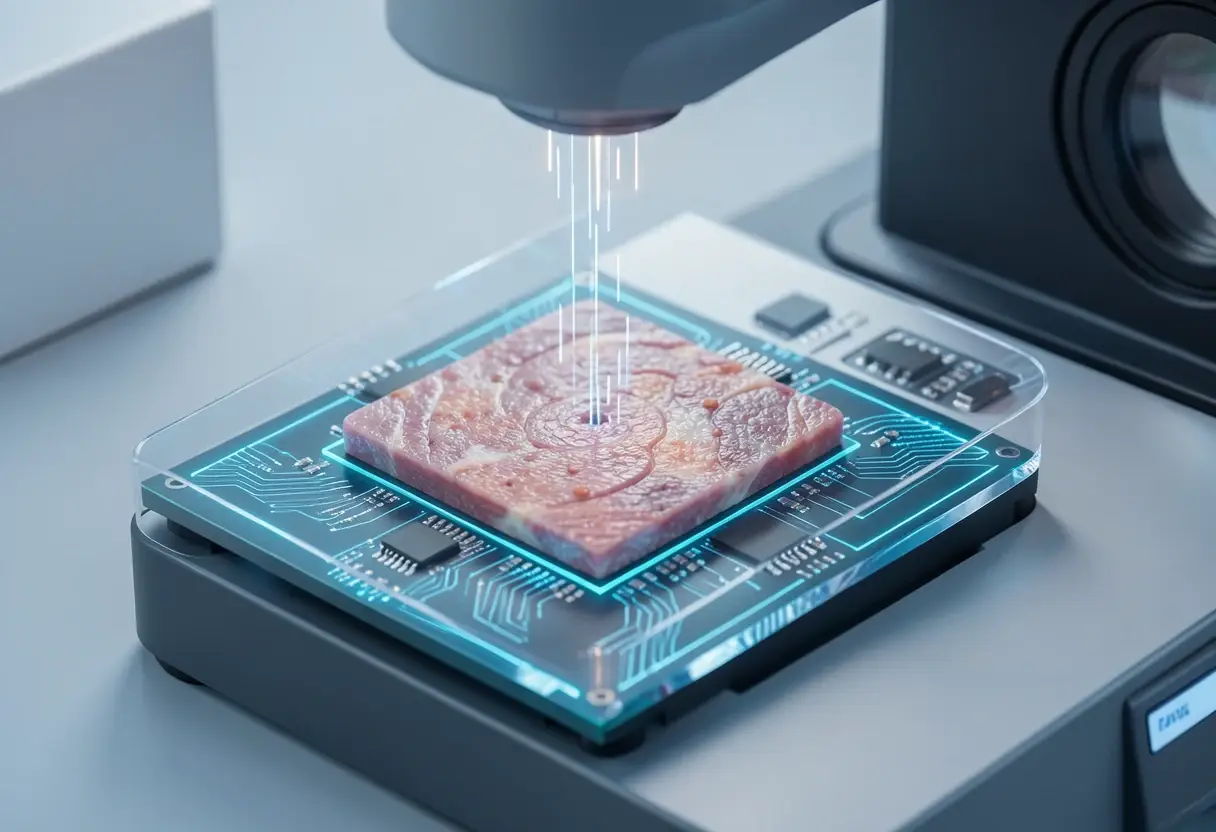
The imec and KU Leuven team moved that control onto an optical phased array, an array of microscopic waveguides etched into a photonic chip. Each channel emits light with an independently tuned phase; coordinating those phases shapes the illumination before it enters tissue and compensates for the measured scattering pattern.
Putting the array on a chip also permits other optical functions to share the platform, potentially reducing system size and complexity relative to a separate liquid-crystal modulator. No chip dimensions or packaged-system specifications were disclosed.
What has the team measured?
The experimental series reported a focus contrast of about 90% of the theoretical maximum after light had been strongly scrambled by the scattering medium. The authors described the recovered focus as almost maximum efficiency. The array also operated at about 5,000 corrections per second, which they described as substantially faster than conventional liquid-crystal modulation, although they published no numerical speed ratio.
These are measured laboratory results, not a full imaging specification. The disclosed test details do not specify the optical wavelength, sample tissue, penetration depth, focal spot size, optical power, repeatability, or operating environment. They therefore do not establish how far the method can image in vivo, nor whether 90% contrast and 5,000 corrections per second persist under those conditions.
Which applications could follow?
Neuroscience is a direct candidate because researchers use optical methods to study brain activity but face a depth limit. Better focus could make processes deeper in tissue observable without abandoning optical resolution. Biomedical diagnostics could benefit from higher contrast and sensitivity, although the research does not report disease detection, diagnostic accuracy, clinical sensitivity, specificity, or a regulatory validation.
Multimode optical fiber offers a different route. Telecom fibers carry large data volumes, but their small diameter also makes them candidate ultrathin imaging probes for body locations that conventional instruments cannot reach.
Many propagating paths distort the image; shaping the input with an optical phased array could reconstruct a sharp focus at the fiber output and support high-resolution imaging through a hair-thin probe, reducing the invasiveness of endoscopy. This remains an anticipated application.
What must improve before deployment?
Development now targets higher speed, greater scalability, better robustness and more on-chip functionality. The team also seeks lower system complexity and easier deployment in practical settings. These work items place the technology between proof of concept and broader application; the development description does not announce a commercial instrument, an in vivo trial, a calibration procedure, or a compliance standard.
Who is developing the platform?
- Pol Van Dorpe is an imec fellow in the health sector and a part-time KU Leuven physics associate professor. He earned a KU Leuven PhD in 2006 for spintronics, has worked in spintronics, plasmonics, biophotonics and single-molecule sensing, and became an imec fellow in 2020. His work targets life-science uses of integrated photonics and single-molecule electronic and ionic devices.
- Filip Milojković is an imec researcher developing integrated photonics for biomedical uses, including wavefront shaping in microscopy. He earned a University of Belgrade BSc in 2019 and an MSc in photonics from Ghent University and VUB in 2021, began doctoral research at imec and KU Leuven in 2021, and received a KU Leuven PhD in physics in 2026.
- Niels Verellen is imec's scientific director for exploratory photonics and a KU Leuven quantum solid-state physics fellow. His work targets application enablers for life sciences and quantum sensing. He earned an MSc in 2007 and a PhD in 2011, received the Umicore Scientific Award in 2012, and holds ERC Starting and Proof of Concept grants for an integrated-photonics high-throughput microscopy platform.
Which papers report the results?
- F. Milojković et al., ACS Photonics 11(10), 4288–4296 (2024); DOI: https://doi.org/10.1021/acsphotonics.4c01228.
- F. Milojković et al., ACS Photonics 12(11), 6335–6342 (2025); DOI: https://doi.org/10.1021/acsphotonics.5c01814.
- F. Milojković et al., Nanophotonics 14, 3255–3264 (2025); DOI: https://doi.org/10.1515/nanoph-2025-0273.
For adoption, the decisive test is whether the team can reproduce 90% focus contrast and 5,000 corrections per second in living tissue at specified wavelengths, powers and depths, then package the chip without sacrificing stability.
Can the platform preserve that performance in breathing, perfused tissue and in a packaged system? Until those tests exist, the result remains a laboratory demonstration of fast wavefront control, not a qualified deep-tissue imager.
via doi.org (Original)
Filed under
- wavefront-shaping
- optical-phased-array
- photonic-chip
- microscopy
- scattering-media
More from Nathan Brooks
Show full bio
Staff writer covering people, launches and day-to-day practice in Instruments, test and measurement at Testbench Report.
78 articles